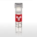
Thermo Fisher Scientific CD63 Monoclonal Antibody (NVG-2), PE-Cyanine7, eBioscience
✨AI 추천 연관 상품
AI가 분석한 이 상품과 연관된 추천 상품들을 확인해보세요
연관 상품을 찾고 있습니다...
Applications
Tested Dilution
Publications
Flow Cytometry (Flow)
0.125 µg/test
View 1 publication 1 publication
Product Specifications
Published species
Not Applicable
Host/Isotype
Rat / IgG2a, kappa
Recommended Isotype Control
Rat IgG2a kappa Isotype Control (eBR2a), PE-Cyanine7, eBioscience™
Class
Monoclonal
Type
Antibody
Clone
NVG-2
Conjugate
PE-Cyanine7 PE-Cyanine7 PE-Cyanine7
View additional formats
Excitation/Emission Max
569/780 nm View spectra 
Form
Liquid
Concentration
0.2 mg/mL
Storage conditions
4° C, store in dark, DO NOT FREEZE!
Shipping conditions
Wet ice
RRID
AB_2573355
Product Specific Information
Description: The monoclonal antibody NVG-2 reacts with mouse CD63, also known as Lysosomal-Associated Membrane Protein 3 (LAMP-3) or tetraspanin 30 (TSPN30), a member of tetraspanin family of proteins characterized by four transmembrane domains. CD63 is expressed on a variety of cell types of hematopoietic lineage, e.g., granulocytes, B lymphocytes, platelets, as well as cells of non-hematopoietic origin. It can be found on the cell membrane, late endocytic vesicles, lysosomes, exosomes, and other specialized granules. On the cell surface, CD63 has been shown to interact with various proteins forming tetraspanin-enriched microdomains (TEM). Its high expression on the cell membrane may be indicative of cell activation, hence, CD63 is often used as an activation marker for basophils, platelets and other cells.
Applications Reported: This NVG-2 antibody has been reported for use in intracellular staining followed by flow cytometric analysis.
Applications Tested: This NVG-2 antibody has been tested by intracellular staining followed by flow cytometric analysis of mouse resident peritoneal exudate cells using the Intracellular Fixation & Permeabilization Buffer Set (Product # 88-8824-00) and protocol. Please refer to BestProtocols®: Protocol A: Two step protocol for (cytoplasmic) intracellular proteins located under the Resources Tab online. This can be used at less than or equal to 0.125 µg per test. A test is defined as the amount (µg) of antibody that will stain a cell sample in a final volume of 100 µL. Cell number should be determined empirically but can range from 10^5 to 10^8 cells/test. It is recommended that the antibody be carefully titrated for optimal performance in the assay of interest.
Light sensitivity: This tandem dye is sensitive to photo-induced oxidation. Please protect this vial and stained samples from light.
Fixation: Samples can be stored in IC Fixation Buffer (Product # 00-822-49) (100 µL of cell sample + 100 µL of IC Fixation Buffer) or 1-step Fix/Lyse Solution (Product # 00-5333-54) for up to 3 days in the dark at 4°C with minimal impact on brightness and FRET efficiency/compensation. Some generalizations regarding fluorophore performance after fixation can be made, but clone specific performance should be determined empirically.
Excitation: 488-561 nm; Emission: 775 nm; Laser: Blue Laser, Green Laser, Yellow-Green Laser.
Filtration: 0.2 µm post-manufacturing filtered.
Target Information
CD63 (LAMP-3, lysosome-associated membrane protein-3), a glycoprotein of tetraspanin family, is present in late endosomes, lysosomes and secretory vesicles of various cell types. CD63 is also present in the plasma membrane, usually following cell activation. Hence, CD63 has become a widely used basophil activation marker. In mast cells, however, CD63 exposition does not need their activation. CD63 interacts with integrins and affects phagocytosis and cell migration, it is also involved in H/K-ATPase trafficking regulation of ROMK1 channels. CD63 also serves as a T-cell costimulation molecule. Expression of CD63 can be used for predicting the prognosis in earlier stages of carcinomas. CD63 is expressed on activated platelets, and is a lysosomal membrane glycoprotein that is translocated to plasma membrane after platelet activation. CD63 is also present in monocytes and macrophages and is weakly expressed on granulocytes, B, and T cells. CD63 is identical to the melanoma-associated antigen which is ME491 and to the platelet antigen PTLGP40. Diseases associated with CD63 dysfunction include melanoma and Hermansky-Pudlak Syndrome.
For Research Use Only. Not for use in diagnostic procedures. Not for resale without express authorization.
🏷️Thermo Fisher Scientific 상품 둘러보기
동일 브랜드의 다른 상품들을 확인해보세요

Thermo Fisher Scientific
Thermo Fisher Scientific CD62L (L-Selectin) Monoclonal Antibody (MEL-14), PE-Cyanine7, eBioscience
215,100원

Thermo Fisher Scientific
Thermo Fisher Scientific CD66a (CEACAM1) Monoclonal Antibody (CC1), PE-Cyanine7, eBioscience
383,200원

Thermo Fisher Scientific
Thermo Fisher Scientific CD63 Monoclonal Antibody (NVG-2), PE-Cyanine7, eBioscience
228,900원

Thermo Fisher Scientific
Thermo Fisher Scientific CD57 Monoclonal Antibody (TB01 (TBO1)), PE-Cyanine7, eBioscience
542,600원

Thermo Fisher Scientific
Thermo Fisher Scientific CD62L (L-Selectin) Monoclonal Antibody (DREG-56 (DREG56)), PE-Cyanine7, eBioscience
266,900원
배송/결제/교환/반품 안내
배송 정보
| 기본 배송비 |
| 교환/반품 배송비 |
|
|---|---|---|---|
| 착불 배송비 |
| ||
| 교환/반품 배송비 |
| ||
결제 및 환불 안내
| 결제수단 |
|
|---|---|
| 취소 |
|
| 반품 |
|
| 환급 |
|
교환 및 반품 접수
| 교환 및 반품 접수 기한 |
|
|---|---|
| 교환 및 반품 접수가 가능한 경우 |
|
| 교환 및 반품 접수가 불가능한 경우 |
|
교환 및 반품 신청
| 교환 절차 |
|
|---|---|
| 반품 절차 |
|
