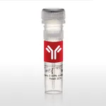
Thermo Fisher Scientific TCR V beta 11 Monoclonal Antibody (RR3-15), PerCP-eFluor 710, eBioscience
다른 상품 둘러보기
Applications
Tested Dilution
Publications
Flow Cytometry (Flow)
0.125 µg/test
Product Specifications
Species Reactivity
Mouse
Host/Isotype
Rat / IgG2b, kappa
Recommended Isotype Control
Rat IgG2b kappa Isotype Control (eB149/10H5), PerCP-eFluor™ 710, eBioscience™
Class
Monoclonal
Type
Antibody
Clone
RR3-15
Conjugate
PerCP-eFluor™ 710 PerCP-eFluor™ 710 PerCP-eFluor™ 710
View additional formats
Excitation/Emission Max
482/708 nm View spectra 
Form
Liquid
Concentration
0.2 mg/mL
Purification
Affinity chromatography
Storage buffer
PBS, pH 7.2
Contains
0.09% sodium azide
Storage conditions
4° C, store in dark, DO NOT FREEZE!
Shipping conditions
Ambient (domestic); Wet ice (international)
RRID
AB_10853822
Product Specific Information
Description: This RR3-15 monoclonal antibody reacts with the mouse T-cell receptor (TCR) V beta 11 chain. Composed of an alpha and beta chain, TCR specificity is typically determined by Va, Ja, Vb, Db, and Jb gene rearrangement. The RR3-15 antibody recognizes the V beta 11 chain on T cells from mouse strains with the b haplotype in the Tcrb gene complex, including C57BL/6 and B10, but is absent in mice with the a haplotype, such as C57L, SJL, and SWR. In addition, reports indicate that Vbeta11+ T cells are eliminated in mice bearing the MHC Class II I-E molecule. Finally, V beta 11+CD4+ T cells have been associated with graft-versus-host disease (GVHD) severity.
The RR3-15 antibody has been reported to activate V beta 11+ T cells.
Applications Reported: The RR3-15 antibody has been reported for use in flow cytometric analysis.
Applications Tested: This RR3-15 antibody has been tested by flow cytometric analysis of mouse lymph node cells. This can be used at less than or equal to 0.125 µg per test. A test is defined as the amount (µg) of antibody that will stain a cell sample in a final volume of 100 µL. Cell number should be determined empirically but can range from 10^5 to 10^8 cells/test. It is recommended that the antibody be carefully titrated for optimal performance in the assay of interest.
PerCP-eFluor® 710 emits at 710 nm and is excited with the blue laser (488 nm); it can be used in place of PerCP-Cyanine5.5. We recommend using a 710/50 bandpass filter, however, the 695/40 bandpass filter is an acceptable alternative. Please make sure that your instrument is capable of detecting this fluorochrome.
Light sensitivity: This tandem dye is sensitive to photo-induced oxidation. Please protect this vial and stained samples from light.
Fixation: Samples can be stored in IC Fixation Buffer (Product # 00-8222) (100 µL of cell sample + 100 µL of IC Fixation Buffer) or 1-step Fix/Lyse Solution (Product # 00-5333) for up to 3 days in the dark at 4°C with minimal impact on brightness and FRET efficiency/compensation. Some generalizations regarding fluorophore performance after fixation can be made, but clone specific performance should be determined empirically.
Excitation: 488 nm; Emission: 710 nm; Laser: Blue Laser.
Filtration: 0.2 µm post-manufacturing filtered.
Target Information
The ability of T cell receptors (TCR) to discriminate foreign from self-peptides presented by major histocompatibility complex (MHC) class II molecules is essential for an effective adaptive immune response. TCR recognition of self-peptides has been linked to autoimmune disease. Mutant self-peptides have been associated with tumors. Engagement of TCRs by a family of bacterial toxins know as superantigens has been responsible for toxic shock syndrome. Autoantibodies to V beta segments of T cell receptors have been isolated from patients with rheumatoid arthritis (RA) and systemic lupus erythematosus (SLE). The autoantibodies block TH1-mediated inflammatory autodestructive reactions and are believed to be a method by which the immune system compensates for disease. Most human T cells express the TCR alpha-beta and either CD4 or CD8 molecule (single positive, SP). A small number of T cells lack both CD4 and CD8 (double negative, DN). Increased percentages of alpha-beta DN T cells have been identified in some autoimmune and immunodeficiency disorders. Gamma-delta T cells are primarily found within the epithelium. They show less TCR diversity and recognize antigens differently than alpha-beta T cells. Subsets of gamma-delta T cells have shown antitumor and immunoregulatory activity.
For Research Use Only. Not for use in diagnostic procedures. Not for resale without express authorization.
배송/결제/교환/반품 안내
배송 정보
| 기본 배송비 |
| 교환/반품 배송비 |
|
|---|---|---|---|
| 착불 배송비 |
| ||
| 교환/반품 배송비 |
| ||
결제 및 환불 안내
| 결제수단 |
|
|---|---|
| 취소 |
|
| 반품 |
|
| 환급 |
|
교환 및 반품 접수
| 교환 및 반품 접수 기한 |
|
|---|---|
| 교환 및 반품 접수가 가능한 경우 |
|
| 교환 및 반품 접수가 불가능한 경우 |
|
교환 및 반품 신청
| 교환 절차 |
|
|---|---|
| 반품 절차 |
|

