Countstar Castor S1
상품 한눈에 보기
고속 3D 오가노이드 분석을 위한 AI 기반 이미지 분석 시스템. Z-stack 기능으로 입체적 구조 분석 가능. 듀얼 형광 채널과 고감도 냉각 카메라 탑재로 정밀한 세포 관찰 지원. FDA 21 CFR Part 11 규정 준수 및 자동화 플랫폼 통합 가능.
브랜드: Countstar
✨AI 추천 연관 상품
AI가 분석한 이 상품과 연관된 추천 상품들을 확인해보세요
연관 상품을 찾고 있습니다...
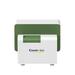
Countstar Castor S1
High-throughput Intelligent 3D Cell Analyzer
Countstar Castor S1은 오가노이드 기반 연구 및 치료 개발을 위한 고속 3D 이미지 분석 플랫폼입니다.
AI 기반 이미지 인식 알고리즘과 Z-stack 기능을 통해 수천 개의 오가노이드를 동시에 정밀 분석할 수 있습니다.
고감도 냉각 카메라와 듀얼 형광 채널을 탑재하여 고해상도 이미지를 확보하며, 직관적인 소프트웨어로 대용량 데이터를 효율적으로 처리합니다.
FDA 21 CFR Part 11 규정을 완벽히 준수하며, 자동화 플랫폼과의 통합이 가능합니다.
Core Advantages
- 고정밀 광학 시스템과 Peltier 냉각 10MP CMOS 카메라로 선명한 이미지 획득
- 듀얼 형광 채널 (Ex480/30nm–Em535/40nm; Ex540/25nm–Em620/60nm)로 다양한 형광 분석 가능
- Z-stack 3D 이미지 및 프로젝션 기능으로 입체적 오가노이드 분석
- AI 기반 이미지 분석 알고리즘으로 자동화된 형태 및 크기 분석
- 6~384 well plate, petri dish, culture flask, slide 등 다양한 소모품 호환
- FDA 21 CFR Part 11 준수 소프트웨어로 데이터 무결성 보장
- 자동화 시스템과 연동 가능하여 고속 처리 지원
Applications Overview
Countstar Castor S1은 오가노이드 배양 품질, 약물 반응 테스트, 오가노이드 생존율 및 면역세포 공배양 독성 분석 등 다양한 고속 오가노이드 기반 실험에 최적화되어 있습니다.
Selected Applications, Results, and Graphics
I. Automatic Analysis of Organoid Culture Quality
- Z-stacking 및 AI 알고리즘으로 개별 오가노이드의 형태, 크기, 밀도 분석
- 배양 기간 동안 오가노이드 성장 추적 및 정량화
II. Growth Monitoring of Cancer-derived Organoids
- 전체 well 영역을 고해상도로 촬영하여 성장 과정의 형태학적 변화 분석
- AI 기반 자동 데이터 처리로 높은 정확도 제공
III. Label-free Organoid Drug Response Test
- 형광 염색 없이 약물 반응을 정량적으로 평가
- 생존 오가노이드와 사멸 오가노이드 비율 분석
IV. Organoid Fluorescence Viability Analysis
- AO/PI 형광 염색을 통한 생존율 및 사멸율 분석
- Z-axis 스캔을 통한 세포 분포 및 약물 침투 평가
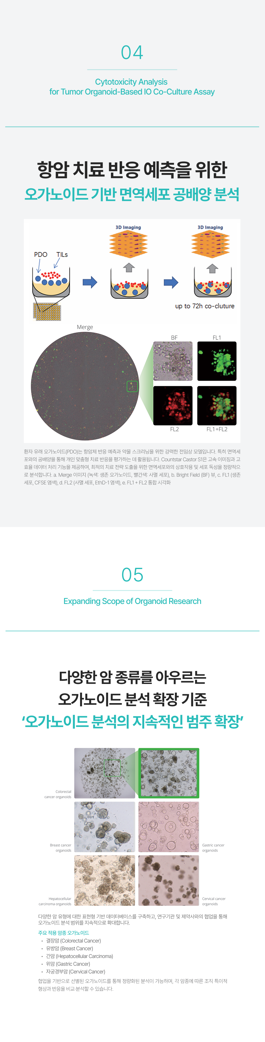
V. Cytotoxicity Analysis for Tumor Organoid-Based IO Co-Culture Assay
- 면역세포와 오가노이드 공배양 분석으로 항암 반응 예측
- FL1/FL2 형광 채널을 이용한 생존 및 사멸 세포 구분
기술 사양 (Technical Specifications)
| 항목 | 내용 |
|---|---|
| 모델명 | Countstar Castor S1 |
| 분석 방식 | Bright Field & Fluorescence Imaging with Z-stack |
| 카메라 | 10MP Peltier-cooled CMOS |
| 배율 | 4.0x / 10.0x 고NA Objective |
| 형광 채널 | Green (480/30–535/40 nm), Red (540/25–620/60 nm) |
| 지원 플레이트 | 6–384 well plate, Petri dish, Flask, Slide |
| 소프트웨어 | AI 기반 이미지 분석, FDA 21 CFR Part 11 준수 |
| 자동화 통합 | 가능 (로봇 및 데이터 관리 시스템 연동) |
이미지 OCR 통합 내용
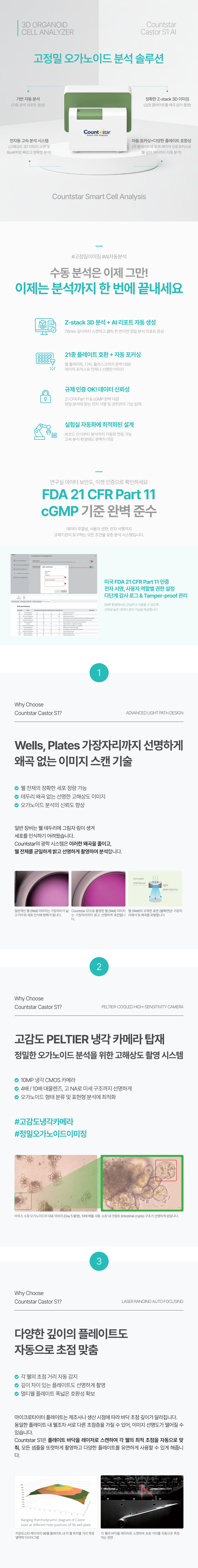
3D ORGANOID CELL ANALYZER – Countstar Castor S1 AI
- AI 기반 오가노이드 인식 및 분류
- Z-stack 3D 분석 및 자동 리포트 생성
- 다양한 플레이트 포맷 지원
- FDA 21 CFR Part 11 및 cGMP 기준 완벽 준수
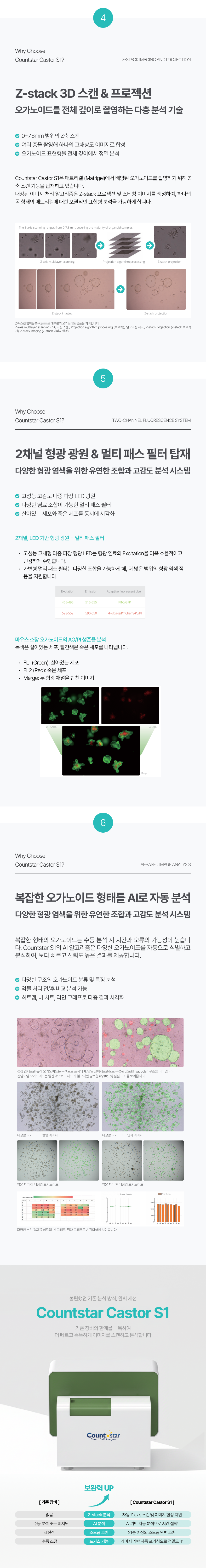
Z-stack 3D 스캔 & 프로젝션
- 0.7μm 단위 Z-스캔으로 오가노이드 전체 깊이 촬영
- 프로젝션 이미지 기반 분석으로 입체적 구조 파악
듀얼 형광 채널
| 채널 | Excitation | Emission | 대표 염색 |
|---|---|---|---|
| Channel 1 | 480 nm | 530 nm | FITC, GFP |
| Channel 2 | 535 nm | 617 nm | PI, RFP |
주요 적용 분야
| 분야 | 설명 |
|---|---|
| 오가노이드 품질 분석 | 크기, 밀도, 형태 정량 분석 |
| 약물 반응 분석 | 약물 처리 전후 변화 비교 |
| 면역세포 상호작용 분석 | 공배양 시스템 내 상호작용 평가 |
| 암 오가노이드 마이그레이션 | 이동 및 침윤 능력 분석 |
Cytotoxicity & Cancer Organoid Expansion
- 다양한 암종 오가노이드 분석 (결장암, 유방암, 간암, 위암, 자궁경부암)
- 면역세포 공배양 기반 독성 및 반응 평가
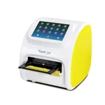
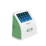
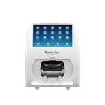
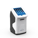
제품 이미지


배송/결제/교환/반품 안내
배송 정보
| 기본 배송비 |
| 교환/반품 배송비 |
|
|---|---|---|---|
| 착불 배송비 |
| ||
| 교환/반품 배송비 |
| ||
결제 및 환불 안내
| 결제수단 |
|
|---|---|
| 취소 |
|
| 반품 |
|
| 환급 |
|
교환 및 반품 접수
| 교환 및 반품 접수 기한 |
|
|---|---|
| 교환 및 반품 접수가 가능한 경우 |
|
| 교환 및 반품 접수가 불가능한 경우 |
|
교환 및 반품 신청
| 교환 절차 |
|
|---|---|
| 반품 절차 |
|
문의 0
로그인 후 문의를 할 수 있습니다.