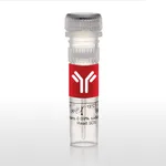
Thermo Fisher Scientific CD66a (CEACAM1) Monoclonal Antibody (CC1), Super Bright 436, eBioscience
다른 상품 둘러보기
Applications
Tested Dilution
Publications
Western Blot (WB)
-
View 1 publication 1 publication
Flow Cytometry (Flow)
0.125 µg/test
View 1 publication 1 publication
Product Specifications
Species Reactivity
Mouse
Published species
Not Applicable
Host/Isotype
Mouse / IgG1, kappa
Recommended Isotype Control
Mouse IgG1 kappa Isotype Control (P3.6.2.8.1), Super Bright™ 436, eBioscience™
Class
Monoclonal
Type
Antibody
Clone
CC1
Conjugate
Super Bright™ 436 Super Bright™ 436 Super Bright™ 436
View additional formats
Excitation/Emission Max
413/431 nm View spectra 
Form
Liquid
Concentration
0.2 mg/mL
Purification
Affinity chromatography
Storage buffer
PBS, pH 7.2, with BSA
Contains
0.09% sodium azide
Storage conditions
4° C, store in dark, DO NOT FREEZE!
Shipping conditions
Ambient (domestic); Wet ice (international)
RRID
AB_2784813
Product Specific Information
Description: The monoclonal antibody CC1 recognizes CD66a, also known as carcinoembryonic antigen-related cell adhesion molecule 1 (CEACAM1), biliary glycoprotein, and BPG. Expression of CD66a is found on brush borders, epithelial, and endothelial cells. In hematopoietic cells expression is found abundantly on B cells, as well as some NKs, monocytes, DCs, and granulocytes. Although low levels of mRNA have been identified in T cells in humans, resting mouse T lymphocytes are not reported to express CD66a, as confirmed by lack of staining with CC1 antibody. In humans, expression levels of CD66a have been used to identify malignancies. CD66a plays a key role as a regulator of BCR activation of B lymphocytes.
An alternate allele, CEACAM1b, is expressed in SJL mice; therefore, CC1 does not stain SJL tissue. The monoclonal CC1 has been shown to block viral infection and also enhance B cell proliferation when combined with IgM crosslinking.
Applications Reported: This CC1 antibody has been reported for use in flow cytometric analysis.
Applications Tested: This CC1 antibody has been tested by flow cytometric analysis of mouse splenocytes. This may be used at less than or equal to 0.125 µg per test. A test is defined as the amount (µg) of antibody that will stain a cell sample in a final volume of 100 µL. Cell number should be determined empirically but can range from 10^5 to 10^8 cells/test. It is recommended that the antibody be carefully titrated for optimal performance in the assay of interest.
Super Bright 436 can be excited with the violet laser line (405 nm) and emits at 436 nm. We recommend using a 450/50 bandpass filter, or equivalent. Please make sure that your instrument is capable of detecting this fluorochrome.
When using two or more Super Bright dye-conjugated antibodies in a staining panel, it is recommended to use Super Bright Complete Staining Buffer (Product # SB-4401) to minimize any non-specific polymer interactions. Please refer to the datasheet for Super Bright Staining Buffer for more information.
Excitation: 405 nm; Emission: 436 nm; Laser: Violet Laser
Super Bright Polymer Dyes are sold under license from Becton, Dickinson and Company.
Target Information
CEACAM1 is a member of the carcinoembryonic antigen (CEA) gene family, which belongs to the immunoglobulin superfamily. Two subgroups of the CEA family, the CEA cell adhesion molecules and the pregnancy-specific glycoproteins, are located within a 1.2 Mb cluster on the long arm of chromosome 19. Eleven pseudogenes of the CEA cell adhesion molecule subgroup are also found in the cluster. The encoded protein was originally described in bile ducts of liver as biliary glycoprotein. Subsequently, it was found to be a cell-cell adhesion molecule detected on leukocytes, epithelia, and endothelia. The encoded protein mediates cell adhesion via homophilic as well as heterophilic binding to other proteins of the subgroup. Multiple cellular activities have been attributed to the encoded protein, including roles in the differentiation and arrangement of tissue three-dimensional structure, angiogenesis, apoptosis, tumor suppression, metastasis, and the modulation of innate and adaptive immune responses. Multiple transcript variants encoding different isoforms have been reported, but the full-length nature of only two has been determined.
For Research Use Only. Not for use in diagnostic procedures. Not for resale without express authorization.
배송/결제/교환/반품 안내
배송 정보
| 기본 배송비 |
| 교환/반품 배송비 |
|
|---|---|---|---|
| 착불 배송비 |
| ||
| 교환/반품 배송비 |
| ||
결제 및 환불 안내
| 결제수단 |
|
|---|---|
| 취소 |
|
| 반품 |
|
| 환급 |
|
교환 및 반품 접수
| 교환 및 반품 접수 기한 |
|
|---|---|
| 교환 및 반품 접수가 가능한 경우 |
|
| 교환 및 반품 접수가 불가능한 경우 |
|
교환 및 반품 신청
| 교환 절차 |
|
|---|---|
| 반품 절차 |
|

