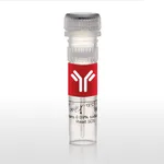
Thermo Fisher Scientific CD8a Monoclonal Antibody (OX8), PE-Cyanine7, eBioscience
✨AI 추천 연관 상품
AI가 분석한 이 상품과 연관된 추천 상품들을 확인해보세요
연관 상품을 찾고 있습니다...
Applications
Tested Dilution
Publications
Flow Cytometry (Flow)
0.25 µg/test
View 10 publications 10 publications
Product Specifications
Species Reactivity
Rat
Published species
Rat
Host/Isotype
Mouse / IgG1, kappa
Recommended Isotype Control
Mouse IgG1 kappa Isotype Control (P3.6.2.8.1), PE-Cyanine7, eBioscience™
Class
Monoclonal
Type
Antibody
Clone
OX8
Conjugate
PE-Cyanine7 PE-Cyanine7 PE-Cyanine7
View additional formats
- Unconjugated
- Alexa Fluor 700
- APC
- eFluor 450
- FITC
- PE
- PerCP-eFluor 710
- Super Bright 436
- Super Bright 600
- Super Bright 780
- Request custom conjugation
Excitation/Emission Max
569/780 nm View spectra 
Form
Liquid
Concentration
0.2 mg/mL
Purification
Affinity chromatography
Storage buffer
PBS, pH 7.2
Contains
0.09% sodium azide
Storage conditions
4° C, store in dark, DO NOT FREEZE!
Shipping conditions
Ambient (domestic); Wet ice (international)
RRID
AB_10548361
Product Specific Information
Description: The OX8 monoclonal antibody reacts with rat CD8 alpha. CD8 alpha is a member of the immunoglobulin superfamily which heterodimerizes with CD8 beta to form the CD8 antigen expressed on CD4+CD8+, double-positive thymocytes, the majority of MHC class I-restricted peripheral T cells, and on some macrophages. CD8 binds to MHC class I expressed on the surface of antigen-presenting cells during antigen presentation, and participates in T-cell receptor signal transduction through association with the kinase Lck.
Applications Reported: This OX8 antibody has been reported for use in flow cytometric analysis.
Applications Tested: This OX8 antibody has been tested by flow cytometric analysis of rat splenocytes. This can be used at less than or equal to 0.25 µg per test. A test is defined as the amount (µg) of antibody that will stain a cell sample in a final volume of 100 µL. Cell number should be determined empirically but can range from 10^5 to 10^8 cells/test. It is recommended that the antibody be carefully titrated for optimal performance in the assay of interest.
Light sensitivity: This tandem dye is sensitive photo-induced oxidation. Please protect this vial and stained samples from light.
Fixation: Samples can be stored in IC Fixation Buffer (Product # 00-822-49) (100 µL cell sample + 100 µL IC Fixation Buffer) or 1-step Fix/Lyse Solution (Product # 00-5333-54) for up to 3 days in the dark at 4°C with minimal impact on brightness and FRET efficiency/compensation. Some generalizations regarding fluorophore performance after fixation can be made, but clone specific performance should be determined empirically.
Excitation: 488-561 nm; Emission: 775 nm; Laser: Blue Laser, Green Laser, Yellow-Green Laser.
Filtration: 0.2 µm post-manufacturing filtered.
Target Information
Cluster of differentiation 8 (CD8), a type I transmembrane glycoprotein of the immunoglobulin family of receptors, plays an integral role in signal transduction, and T cell differentiation and activation. CD8 is predominantly expressed on T cells as a disulfide-linked heterodimer of CD8alpha and CD8beta, where it functions as a co-receptor, along with T cell receptor (TCR), for major histocompatibilty complex class I (MHC-I) molecules; whereas its counterpart, CD4, acts as a co-receptor for MHC-II molecules. CD8 exists on the cell surface, where the CD8alpha chain is essential for binding to MHC-I. CD8 is also expressed on a subset of T cells, NK cells, monocytes and dendritic cells as disulfide-linked homodimers of CD8alpha. Ligation of MHC-I/peptide complexes presented by antigen-presenting cells (APCs), triggers the recruitment of lymphocyte-specific protein tyrosine kinase (Lck), which leads to lymphokine production, motility and cytotoxic T lymphocyte (CTL) activation. Once activated, CTLs play a crucial role in the clearance of pathogens and tumor cells. Differentiation of naive CD8+ T cells into CTLs is strongly enhanced by IL-2, IL-12 and TGF-beta1.
For Research Use Only. Not for use in diagnostic procedures. Not for resale without express authorization.
🏷️Thermo Fisher Scientific 상품 둘러보기
동일 브랜드의 다른 상품들을 확인해보세요

Thermo Fisher Scientific
Thermo Fisher Scientific CD8b Monoclonal Antibody (eBioH35-17.2 (H35-17.2)), PE-Cyanine7, eBioscience
489,800원

Thermo Fisher Scientific
Thermo Fisher Scientific CD8a Monoclonal Antibody (SK1), PE-Cyanine7, eBioscience
255,200원

Thermo Fisher Scientific
Thermo Fisher Scientific CD8a Monoclonal Antibody (OX8), PE-Cyanine7, eBioscience
415,500원

Thermo Fisher Scientific
Thermo Fisher Scientific CD7 Monoclonal Antibody (eBio124-1D1 (124-1D1)), PE-Cyanine7, eBioscience
441,900원

Thermo Fisher Scientific
Thermo Fisher Scientific Mouse G-CSF Recombinant Protein, PeproTech
138,900원
배송/결제/교환/반품 안내
배송 정보
| 기본 배송비 |
| 교환/반품 배송비 |
|
|---|---|---|---|
| 착불 배송비 |
| ||
| 교환/반품 배송비 |
| ||
결제 및 환불 안내
| 결제수단 |
|
|---|---|
| 취소 |
|
| 반품 |
|
| 환급 |
|
교환 및 반품 접수
| 교환 및 반품 접수 기한 |
|
|---|---|
| 교환 및 반품 접수가 가능한 경우 |
|
| 교환 및 반품 접수가 불가능한 경우 |
|
교환 및 반품 신청
| 교환 절차 |
|
|---|---|
| 반품 절차 |
|
