1 / 2
Merck Anti-Tau-1 Antibody, clone PC1C6
상품 한눈에 보기
Tau 단백질의 인산화 상태를 구분하여 인식하는 mouse monoclonal antibody. 알츠하이머병 관련 신경섬유 병변 연구에 적합. 신경세포 축삭 특이적 염색. 세포 배양 및 조직 샘플 모두에 사용 가능.
브랜드: Merck Sigma
✨AI 추천 연관 상품
AI가 분석한 이 상품과 연관된 추천 상품들을 확인해보세요
연관 상품을 찾고 있습니다...
Anti-Tau-1 Antibody, clone PC1C6
개요
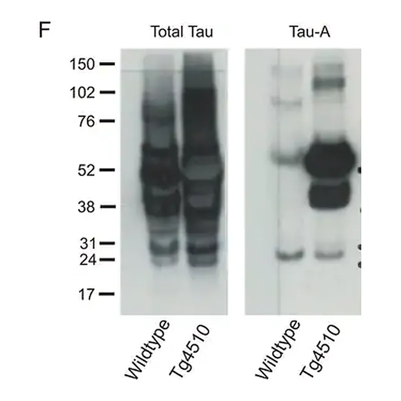
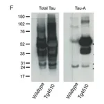
Anti-Tau-1 항체(clone PC1C6)는 Chemicon®에서 유래된 mouse monoclonal antibody로, Tau 단백질을 인식합니다. Tau는 미세소관 안정화에 관여하는 단백질로, 알츠하이머병의 주요 병리적 특징인 paired helical filaments (PHF)의 주요 성분으로 알려져 있습니다.
Tau 단백질의 과인산화(hyperphosphorylation)는 PHF 형성의 핵심 단계로 여겨지며, 총 6종의 Tau isoform이 알려져 있습니다. 이들은 모두 glycogen synthase kinase 3 (GSK3)에 의해 인산화됩니다.
세포 및 세포소기관 특이성
- In situ: Anti-Tau-1은 신경세포의 축삭(axon)에 대해 높은 특이성을 보입니다. 세포체(cell body)나 수상돌기(dendrite), 기타 세포 유형은 염색되지 않습니다.
- In vitro: 배양된 쥐 해마 뉴런에서는 축삭, 세포체, 수상돌기 모두 염색됩니다.
- 인산화 상태 의존성:
- 탈인산화 처리된 시료(알칼리성 인산분해효소 처리)에서는 성상세포(astrocyte), 신경주위 교세포(perineuronal glial cell), 축삭, 세포체, 수상돌기가 모두 염색됩니다.
- 처리하지 않은 시료에서는 축삭만 염색됩니다.
- Anti-Tau-1이 인식하는 epitope은 인산화 부위 근처에 존재하는 것으로 추정됩니다.
제품 정보 요약
| 항목 | 내용 |
|---|---|
| 제품명 | Anti-Tau-1 Antibody, clone PC1C6 |
| 클론명 | PC1C6 |
| 호스트 | Mouse |
| 브랜드 | Chemicon® (Merck Sigma) |
| 인식 대상 | Tau 단백질 (phosphorylation state-dependent) |
| 주요 용도 | Tau 단백질 인산화 연구, 알츠하이머병 관련 연구 |
| 적용 분야 | Immunocytochemistry, Immunohistochemistry, Neuroscience research |
🏷️Merck Sigma 상품 둘러보기
동일 브랜드의 다른 상품들을 확인해보세요
배송/결제/교환/반품 안내
배송 정보
| 기본 배송비 |
| 교환/반품 배송비 |
|
|---|---|---|---|
| 착불 배송비 |
| ||
| 교환/반품 배송비 |
| ||
결제 및 환불 안내
| 결제수단 |
|
|---|---|
| 취소 |
|
| 반품 |
|
| 환급 |
|
교환 및 반품 접수
| 교환 및 반품 접수 기한 |
|
|---|---|
| 교환 및 반품 접수가 가능한 경우 |
|
| 교환 및 반품 접수가 불가능한 경우 |
|
교환 및 반품 신청
| 교환 절차 |
|
|---|---|
| 반품 절차 |
|
문의 0
로그인 후 문의를 할 수 있습니다.