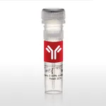
Thermo Fisher Scientific CD273 (B7-DC) Monoclonal Antibody (122), Super Bright 436, eBioscience
✨AI 추천 연관 상품
AI가 분석한 이 상품과 연관된 추천 상품들을 확인해보세요
연관 상품을 찾고 있습니다...
Applications
Tested Dilution
Publications
Flow Cytometry (Flow)
1.0 µg/test
Product Specifications
Species Reactivity
Mouse
Host/Isotype
Rat / IgG2a, kappa
Recommended Isotype Control
Rat IgG2a kappa Isotype Control (eBR2a), Super Bright™ 436, eBioscience™
Class
Monoclonal
Type
Antibody
Clone
122
Conjugate
Super Bright™ 436 Super Bright™ 436 Super Bright™ 436
View additional formats
Excitation/Emission Max
413/431 nm View spectra 
Form
Liquid
Concentration
0.2 mg/mL
Purification
Affinity chromatography
Storage buffer
PBS, pH 7.2, with BSA
Contains
0.09% sodium azide
Storage conditions
4° C, store in dark, DO NOT FREEZE!
Shipping conditions
Ambient (domestic); Wet ice (international)
RRID
AB_2688153
Product Specific Information
Description: The 122 monoclonal antibody reacts with mouse B7-DC, also known as PD-L2. B7-DC, a member of the B7 family, has a predicted molecular weight of approximately 25 kDa and belongs to the Ig superfamily. The mouse B7-DC has a short cytoplasmic tail (4aa). B7-DC is primarily expressed by subpopulations of dendritic cells and monocytes/macrophages in the mouse. Although B7-DC has structural and sequence similarities to the B7 family, it does not bind CD28/CTLA-4, rather it is a ligand for PD-1. The interactions between PD-1 and B7-DC/PD-L2 have been reported to be involved in costimulation or suppression of T cell proliferation depending on state of cellular activation. 122 has been demonstrated to block binding of TY25 (Product # 14-5986), another mAb specific for mouse B7-DC.
Applications Reported: This 122 antibody has been reported for use in flow cytometric analysis.
Applications Tested: This 122 antibody has been tested by flow cytometric analysis of bone marrow derived dendritic cells. This may be used at less than or equal to 1.0 µg per test. A test is defined as the amount (µg) of antibody that will stain a cell sample in a final volume of 100 µL. Cell number should be determined empirically but can range from 10^5 to 10^8 cells/test. It is recommended that the antibody be carefully titrated for optimal performance in the assay of interest.
Super Bright 436 can be excited with the violet laser line (405 nm) and emits at 436 nm. We recommend using a 450/50 bandpass filter, or equivalent. Please make sure that your instrument is capable of detecting this fluorochrome.
When using two or more Super Bright dye-conjugated antibodies in a staining panel, it is recommended to use Super Bright Complete Staining Buffer (Product # SB-4401) to minimize any non-specific polymer interactions. Please refer to the datasheet for Super Bright Staining Buffer for more information.
Excitation: 405 nm; Emission: 436 nm; Laser: Violet Laser
Super Bright Polymer Dyes are sold under license from Becton, Dickinson and Company.
Target Information
Programmed death-ligand 2 (PD-L2), or B7-DC, is a member of the B7 ligand family within the immunoglobulin superfamily that, along with programmed death-ligand 1 (PD-L1), acts as a ligand for programmed cell death protein 1 (PD-1). Though expressed primarily in dendritic cells, PD-L2 expression can be induced on a wide variety of immune and non-immune cells depending on the microenvironment. PD-L2 expression is particularly upregulated in the presence of Th2 cytokine, IL-4, as well as Th1 cytokines, TNF-alpha and IFN-gamma to a lesser degree. While generally expressed at lower levels compared to PD-L1, PD-L2 demonstrates a 2 to 6 times higher relative affinity to PD-1 than PD-L1. PD-1 and its ligands are referred to as inhibitory immune checkpoint molecules in that they provide useful negative feedback during physiological homeostasis. Ligation of PD-L2 or PD-L1 inhibits activation, proliferation, and cytokine secretion (e.g. IFN-gamma, IL-10) in T cells, ultimately dampening immune response. Conversely, studies have shown that PD-L2 can also stimulate T cell proliferation and cytokine production, even in PD-1-deficient T cells, suggesting additional receptors. Recent studies have concluded that PD-L2 also binds to a second receptor, repulsive guidance molecule b (RGMb), which was originally identified as a receptor for bone morphogenetic proteins (BMPs). RGMb is expressed in the central nervous system, as well as in macrophages, however, its role in immunity is only beginning to emerge. Interaction between PD-L2 and RGMb regulates the development of respiratory tolerance in the lung through BMP and/or neogenin signaling pathways. The naturally occurring human PD-L2 monomer consists of a 201-amino-acid extracellular domain, a 21-amino-acid transmembrane domain, and a 32-amino-acid cytoplasmic domain.
For Research Use Only. Not for use in diagnostic procedures. Not for resale without express authorization.
🏷️Thermo Fisher Scientific 상품 둘러보기
동일 브랜드의 다른 상품들을 확인해보세요

Thermo Fisher Scientific
Thermo Fisher Scientific CD279 (PD-1) Monoclonal Antibody (RMP1-30), Super Bright 436, eBioscience
653,000원

Thermo Fisher Scientific
Thermo Fisher Scientific CD235a (Glycophorin A) Monoclonal Antibody (HIR2 (GA-R2)), Super Bright 436, eBioscience
628,600원

Thermo Fisher Scientific
Thermo Fisher Scientific CD273 (B7-DC) Monoclonal Antibody (122), Super Bright 436, eBioscience
1,950,200원

Thermo Fisher Scientific
Thermo Fisher Scientific Nalgene Lab Notebooks with PolyPaper Pages
60,400원

Thermo Fisher Scientific
Thermo Fisher Scientific Nalgene Lab Notebooks with PolyPaper Pages
362,000원
배송/결제/교환/반품 안내
배송 정보
| 기본 배송비 |
| 교환/반품 배송비 |
|
|---|---|---|---|
| 착불 배송비 |
| ||
| 교환/반품 배송비 |
| ||
결제 및 환불 안내
| 결제수단 |
|
|---|---|
| 취소 |
|
| 반품 |
|
| 환급 |
|
교환 및 반품 접수
| 교환 및 반품 접수 기한 |
|
|---|---|
| 교환 및 반품 접수가 가능한 경우 |
|
| 교환 및 반품 접수가 불가능한 경우 |
|
교환 및 반품 신청
| 교환 절차 |
|
|---|---|
| 반품 절차 |
|
