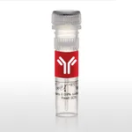
Thermo Fisher Scientific Arginase 1 Monoclonal Antibody (A1exF5), Brilliant Violet 421, eBioscience
✨AI 추천 연관 상품
AI가 분석한 이 상품과 연관된 추천 상품들을 확인해보세요
연관 상품을 찾고 있습니다...
Applications
Tested Dilution
Publications
Flow Cytometry (Flow)
1.0 µg/test
Product Specifications
Species Reactivity
Human, Mouse
Host/Isotype
Rat / IgG2a, kappa
Recommended Isotype Control
Rat IgG2a kappa Isotype Control (eBR2a), Brilliant Violet™ 421, eBioscience™
Class
Monoclonal
Type
Antibody
Clone
A1exF5
Immunogen
E.coli-derived recombinant mouse Arginase 1
Conjugate
Brilliant Violet™ 421 Brilliant Violet™ 421 Brilliant Violet™ 421
View additional formats
- Alexa Fluor 488
- Alexa Fluor 700
- APC
- Brilliant UV 805
- Brilliant Violet 480
- Brilliant Violet 650
- Brilliant Violet 711
- eFluor 450
- PE
- PE-Cyanine7
- PerCP-eFluor 710
- Request custom conjugation
Excitation/Emission Max
406/423 nm View spectra 
Form
Liquid
Concentration
0.2 mg/mL
Purification
Affinity chromatography
Storage buffer
PBS, pH 7.2, with BSA
Contains
0.09% sodium azide
Storage conditions
4° C, store in dark, DO NOT FREEZE!
Shipping conditions
Wet ice
RRID
AB_3074055
Product Specific Information
Description: The monoclonal antibody A1exF5 recognizes both human and mouse Arginase 1, a cytosolic enzyme (Arg1). This A1exF5 clone is compatible with both, the standard intracellular protocols, and the Foxp3/Transcription Factor Staining Buffer Set.
Applications Reported: This A1exF5 antibody has been reported for use in intracellular staining followed by flow cytometric analysis.
Applications Tested: This A1exF5 antibody has been tested by intracellular staining followed by flow cytometric analysis of Normal human lysed whole blood cells using the Intracellular Fixation & Permeabilization Buffer Set (Product # 88-8824-00) and protocol. Please refer to Staining Intracellular Antigens for Flow Cytometry, Protocol A: Two step protocol for intracellular (cytoplasmic) proteins located at Flow Protocols. This may be used at less than or equal to 1.0 µg per test. A test is defined as the amount (µg) of antibody that will stain a cell sample in a final volume of 100 µL. Cell number should be determined empirically but can range from 10^5 to 10^8 cells/test. It is recommended that the antibody be carefully titrated for optimal performance in the assay of interest.
Brilliant Violet™ 421 (BV421) is a dye that emits at 423 nm and is intended for use on cytometers equipped with a violet (405 nm) laser. Please make sure that your instrument is capable of detecting this fluorochrome.
When using two or more Super Bright, Brilliant Violet™, Brilliant Ultra Violet™, or other polymer dye-conjugated antibodies in a staining panel, it is recommended to use Super Bright Complete Staining Buffer (Product # SB-4401-42) or Brilliant Stain Buffer™ (Product # 00-4409-75) to minimize any non-specific polymer interactions. Please refer to the datasheet for Super Bright Staining Buffer or Brilliant Stain Buffer for more information.
Excitation: 407 nm; Emission: 423 nm; Laser: Violet Laser.
BRILLIANT VIOLET™ is a trademark or registered trademark of Becton, Dickinson and Company or its affiliates, and is used under license. Powered by Sirigen™.
Target Information
Arginase-1 (Arg1) is a 35 kDa enzyme converting L-arginine to urea and L-ornithine, which is the final step in the urea cycle. The resulting polyamines are important for cell proliferation and removal of toxins that arise from protein degradation. By degrading arginine, Arginase 1 deprives NO synthase of its substrate and down-regulates nitric oxide production. In both human and mouse, Arginase 1 is expressed in the liver, neutrophils, myeloid derived suppressor cells (MDSC) and neural stem cells. In human, expression in blood neutrophils but not in CCR3+ granulocytes has been reported. In mice, expression of Arginase 1 is one of the hallmarks of alternatively activated macrophages (M2a). Arginase-1 may be expressed in the myeloid cells infiltrating tumors, and is typically found in the majority of hepatocellular carcinomas. Defects in Arginase 1 are the cause of argininemia, an autosomal recessive disorder characterized by hyperammonemia.
For Research Use Only. Not for use in diagnostic procedures. Not for resale without express authorization.
🏷️Thermo Fisher Scientific 상품 둘러보기
동일 브랜드의 다른 상품들을 확인해보세요

Thermo Fisher Scientific
Thermo Fisher Scientific Armenian Hamster IgG Isotype Control (eBio299Arm), Brilliant Violet 421, eBioscience
681,400원

Thermo Fisher Scientific
Thermo Fisher Scientific Mouse IgG2b kappa Isotype Control (eBMG2b), Brilliant Violet 421, eBioscience
528,900원

Thermo Fisher Scientific
Thermo Fisher Scientific Arginase 1 Monoclonal Antibody (A1exF5), Brilliant Violet 421, eBioscience
286,500원

Thermo Fisher Scientific
Thermo Fisher Scientific Rat IgG1 kappa Isotype Control (eBRG1), Brilliant Violet 421, eBioscience
521,100원

Thermo Fisher Scientific
Thermo Fisher Scientific F4/80 Monoclonal Antibody (BM8), Brilliant Violet 421, eBioscience
592,400원
배송/결제/교환/반품 안내
배송 정보
| 기본 배송비 |
| 교환/반품 배송비 |
|
|---|---|---|---|
| 착불 배송비 |
| ||
| 교환/반품 배송비 |
| ||
결제 및 환불 안내
| 결제수단 |
|
|---|---|
| 취소 |
|
| 반품 |
|
| 환급 |
|
교환 및 반품 접수
| 교환 및 반품 접수 기한 |
|
|---|---|
| 교환 및 반품 접수가 가능한 경우 |
|
| 교환 및 반품 접수가 불가능한 경우 |
|
교환 및 반품 신청
| 교환 절차 |
|
|---|---|
| 반품 절차 |
|
