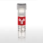
Thermo Fisher Scientific NeuN Recombinant Rabbit Monoclonal Antibody (ZR338), RAbMono
✨AI 추천 연관 상품
AI가 분석한 이 상품과 연관된 추천 상품들을 확인해보세요
연관 상품을 찾고 있습니다...
Applications
Tested Dilution
Publications
Immunohistochemistry (Paraffin) (IHC (P))
Ready-to-use 150-200 µL
Product Specifications
Host/Isotype
Rabbit / IgG
Class
Recombinant Monoclonal
Type
Antibody
Clone
ZR338
Immunogen
Synthetic peptide corresponding to residues within aa 30-60 of human NeuN protein if (typeof window.$mangular === undefined || !window.$mangular) { window.$mangular = {}; } $mangular.antigenJson = \[{targetFamily:NeuN,uniProtId:A6NFN3-1,ncbiNodeId:9606,antigenRange:30-60,antigenLength:312,antigenImageFileName:Z2644RP_NeuN_A6NFN3-1_Rabbit.svg,antigenImageFileNamePDP:Z2644RP_NeuN_A6NFN3-1_Rabbit_PDP.jpeg,sortOrder:1}\]; $mangular.isB2BCMGT = false; $mangular.isEpitopesModalImageMultiSizeEnabled = true;
View immunogen .st0{fill:#FFFFFF;} .st1{fill:#1E8AE7;}
Conjugate
Unconjugated Unconjugated Unconjugated
Form
Liquid
Storage conditions
2-8°C
Shipping conditions
Wet ice
Product Specific Information
This product is diluted and in a ready-to-use formulation.
A recommended positive control tissue for this product is cerebellum, however positive controls are not limited to this tissue type.
The primary antibody is intended for laboratory professional use in the detection of the corresponding protein in formalin-fixed, paraffin-embedded tissue stained in manual qualitative immunohistochemistry (IHC) testing. This antibody is intended to be used after the primary diagnosis of tumor has been made by conventional histopathology using non-immunological histochemical stains.
NeuN antibody specifically recognizes the DNA-binding, neuron-specific protein NeuN, which is present in most CNS and PNS neuronal cell types of all vertebrates tested. NeuN protein distributions are apparently restricted to neuronal nuclei and some proximal neuronal processes in both fetal and adult brain although, some neurons fail to be recognized by NeuN at all ages: INL retinal cells, Cajal-Retzius cells, Purkinje cells, inferior olivary and dentate nucleus neurons, and sympathetic ganglion cells are examples. Immunohistochemically detectable NeuN protein first appears at developmental timepoints that correspond with the withdrawal of the neuron from the cell cycle and/or with the initiation of terminal differentiation of the neuro. Immunoreactivity appears around E9.5 in the mouse neural tube and is extensive throughout the developing nervous system by E12.5. Strong nuclear staining suggests a nuclear regulatory protein function; however, no evidence currently exists as to whether the NeuN protein antigen has a function in the distal cytoplasm or whether it is merely synthesized there before being transported back into the nucleus. No difference between protein isolated from purified nuclei and whole brain extract on immunoblots has been found.
Antibody is used with formalin-fixed and paraffin-embedded sections. Pretreatment of deparaffinized tissue with heat-induced epitope retrieval or enzymatic retrieval is recommended. In general, immunohistochemical (IHC) staining techniques allow for the visualization of antigens via the sequential application of a specific antibody to the antigen (primary antibody), a secondary antibody to the primary antibody (link antibody), an enzyme complex and a chromogenic substrate with interposed washing steps. The enzymatic activation of the chromogen results in a visible reaction product at the antigen site. Results are interpreted using a light microscope and aid in the differential diagnosis of pathophysiological processes, which may or may not be associated with a particular antigen.
A positive tissue control must be run with every staining procedure performed. This tissue may contain both positive and negative staining cells or tissue components and serve as both the positive and negative control tissue. External Positive control materials should be fresh autopsy/biopsy/surgical specimens fixed, processed and embedded as soon as possible in the same manner as the patient sample (s). Positive tissue controls are indicative of correctly prepared tissues and proper staining methods. The tissues used for the external positive control materials should be selected from the patient specimens with well-characterized low levels of the positive target activity that gives weak positive staining. The low level of positivity for external positive controls is designed to ensure detection of subtle changes in the primary antibody sensitivity from instability or problems with the staining methodology. A tissue with weak positive staining is more suitable for optimal quality control and for detecting minor levels of reagent degradation.
Internal or external negative control tissue may be used depending on the guidelines and policies that govern the organization to which the end user belongs to. The variety of cell types present in many tissue sections offers internal negative control sites, but this should be verified by the user. The components that do not stain should demonstrate the absence of specific staining, and provide an indication of non-specific background staining. If specific staining occurs in the negative tissue control sites, results with the patient specimens must be considered invalid.
Target Information
Fox-3, also known as NEUN, HRNBP3, RBFOX3 (RNA binding protein, fox-1) or Fox-1 homolog C, is a 350 amino acid protein that contains one RRM (RNA recognition motif) domain. Localized to both the nucleus and cytoplasm, Fox-3 is suggested to regulate alternative splicing events. Fox-3 is encoded by a gene located on human chromosome 17, which is comprised over 2.5% of the human genome and encodes over 1,200 genes. Two key tumor suppressor genes are associated with chromosome 17, namely, p53 and BRCA1. Tumor suppressor p53 is necessary for maintenance of cellular genetic integrity by moderating cell fate through DNA repair versus cell death. Malfunction or loss of p53 expression is associated with malignant cell growth and LiFraumeni syndrome. Like p53, BRCA1 is directly involved in DNA repair, though specifically it is recognized as a genetic determinant of early onset breast cancer and predisposition to cancers of the ovary, colon, prostate gland and fallopian tubes.
For Research Use Only. Not for use in diagnostic procedures. Not for resale without express authorization.
🏷️Thermo Fisher Scientific 상품 둘러보기
동일 브랜드의 다른 상품들을 확인해보세요

Thermo Fisher Scientific
Thermo Fisher Scientific Olig2 Recombinant Rabbit Monoclonal Antibody (ZR340), RAbMono
544,500원

Thermo Fisher Scientific
Thermo Fisher Scientific Olig2 Recombinant Rabbit Monoclonal Antibody (ZR340), RAbMono
490,800원

Thermo Fisher Scientific
Thermo Fisher Scientific NeuN Recombinant Rabbit Monoclonal Antibody (ZR338), RAbMono
654,400원

Thermo Fisher Scientific
Thermo Fisher Scientific LEF1 Recombinant Rabbit Monoclonal Antibody (ZR336)
621,700원

Thermo Fisher Scientific
Thermo Fisher Scientific NeuN Recombinant Rabbit Monoclonal Antibody (ZR338), RAbMono
461,200원
배송/결제/교환/반품 안내
배송 정보
| 기본 배송비 |
| 교환/반품 배송비 |
|
|---|---|---|---|
| 착불 배송비 |
| ||
| 교환/반품 배송비 |
| ||
결제 및 환불 안내
| 결제수단 |
|
|---|---|
| 취소 |
|
| 반품 |
|
| 환급 |
|
교환 및 반품 접수
| 교환 및 반품 접수 기한 |
|
|---|---|
| 교환 및 반품 접수가 가능한 경우 |
|
| 교환 및 반품 접수가 불가능한 경우 |
|
교환 및 반품 신청
| 교환 절차 |
|
|---|---|
| 반품 절차 |
|
