
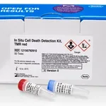
Merck In Situ Cell Death Detection Kit, TMR red
세포 내 DNA 절단을 검출하여 세포 사멸(apoptosis)을 분석하는 TUNEL 기반 키트. TMR red 형광 표지로 시각적 확인 가능. 최대 50회 실험 가능하며, -20°C에서 보관. Roche 제조, Merck Sigma 공급.
✨AI 추천 연관 상품
AI가 분석한 이 상품과 연관된 추천 상품들을 확인해보세요
연관 상품을 찾고 있습니다...
Merck In Situ Cell Death Detection Kit, TMR red
제품 개요
In Situ Cell Death Detection Kit, TMR red는 세포 내 DNA 절단을 검출하여 apoptosis를 분석하는 TUNEL 기반 키트입니다. TMR red 형광 표지를 통해 형광 현미경 또는 유세포분석기로 시각화할 수 있습니다.
사용량
- 최대 50회 실험 가능 (sufficient for ≤50 tests)
품질 등급
- Quality Level: 100
Merck M-Clarity Program
제조사 / 상표명
- Roche
배송 상태
- Dry Ice
저장 온도
- −20°C
원리 (Principle)
이 키트는 apoptosis 초기 단계에서 발생하는 단일 및 이중 가닥 DNA 절단을 검출합니다. 세포를 고정 및 투과 처리한 후, TdT와 TMR-dUTP를 포함하는 TUNEL 반응 혼합물로 처리합니다. TdT는 DNA의 자유 3′-OH 말단에 TMR-dUTP를 부착하며, 이후 형광 현미경 또는 유세포분석기를 통해 손상된 DNA 부위를 시각화할 수 있습니다.
시료 유형 (Sample Material)
- 부유 세포(suspension)
- cytospin 및 cell smear 시료
- 슬라이드 위에 배양된 부착 세포(adherent cells)
- 동결 및 포매(paraffin-embedded) 조직 절편
응용 및 배경
Apoptosis를 확인하는 일반적인 방법에는 DNA 전기영동, 방사성 동위원소 또는 브로모-데옥시유리딘 기반 DNA 절단 분석이 있습니다. 그러나 이러한 방법은 개별 세포 수준의 정보를 제공하지 못합니다.
TUNEL 방법은 DNA 절단의 3′-OH 말단을 표지하여 apoptosis를 정량적이고 시각적으로 검출할 수 있는 민감하고 빠른 방법으로 널리 사용됩니다.
제품 이미지
(이미지 없음)
🏷️Merck Sigma 상품 둘러보기
동일 브랜드의 다른 상품들을 확인해보세요
Merck Sigma
Merck Dispase II (neutral protease, grade II)
Merck Sigma
Merck cOmplete ULTRA Tablets, Mini, EDTA-free, EASYpack Protease Inhibitor Cocktail
Merck Sigma
Merck In Situ Cell Death Detection Kit, TMR red

Merck Sigma
Merck cOmplete, Mini Protease Inhibitor Cocktail
382,200원

Merck Sigma
Merck DNase I recombinant
배송/결제/교환/반품 안내
배송 정보
| 기본 배송비 |
| 교환/반품 배송비 |
|
|---|---|---|---|
| 착불 배송비 |
| ||
| 교환/반품 배송비 |
| ||
결제 및 환불 안내
| 결제수단 |
|
|---|---|
| 취소 |
|
| 반품 |
|
| 환급 |
|
교환 및 반품 접수
| 교환 및 반품 접수 기한 |
|
|---|---|
| 교환 및 반품 접수가 가능한 경우 |
|
| 교환 및 반품 접수가 불가능한 경우 |
|
교환 및 반품 신청
| 교환 절차 |
|
|---|---|
| 반품 절차 |
|