
Thermo Fisher Scientific EVOS M7000 Imaging System, High-Content Analysis Package
완전 자동형 형광 및 투과광 이미징 시스템으로 고해상도 다채널 이미징 지원. Celleste 6.0 소프트웨어 포함으로 고내용 분석 및 데이터 처리 용이. LED 광원과 듀얼 CMOS 카메라로 선명한 이미지 제공. 다양한 배양용기 호환 및 장시간 라이브 셀 이미징 가능.
✨AI 추천 연관 상품
AI가 분석한 이 상품과 연관된 추천 상품들을 확인해보세요
연관 상품을 찾고 있습니다...
Thermo Fisher Scientific EVOS M7000 Imaging System, High-Content Analysis Package
The EVOS™ M7000 Imaging System, High-Content Analysis Package combines a fully automated, inverted, multi-channel fluorescence and transmitted light imaging system with Celleste 6.0 Image Analysis Software, offering optimized protocols for live and fixed cell-based assays. It delivers publication-quality images and data with minimal effort using enhanced autofocus algorithms and automated routines for microwell plate assays, time-lapse imaging, area scanning, and Z-stacks.
주요 특징
- Highly configurable microscope — Five-position objective turret with up to four fluorescence channels plus transmitted light, magnification range 1.25X–100X
- Publication-ready imagery — Autofocus, stitching, tiling, Z-stack, time-lapse imaging, and multichannel capture in a few clicks
- Intuitive acquisition interface — Easily program well-plate scanning, time-lapse experiments, and area scans
- Streamlined image analysis — Celleste 6.0 wizard-driven workflow with preconfigured templates for common assays
Fully Automated Imaging System
The EVOS M7000 integrates precision components for high-quality automated fluorescence imaging. It automates X/Y-stage movement, light cube switching, objective turret rotation, focus, exposure, and dual camera control.
Optional EVOS Onstage Incubator enables long-term live-cell imaging under controlled environmental conditions.
Versatile and Configurable
Compatible with a wide range of objectives (1.25X–100X, achromat, fluorite, apochromat) and LED light cubes for various imaging needs.
Dual monochrome and color CMOS cameras support both fluorescent and chromogenic samples.
Interchangeable vessel holders accommodate slides, multi-well plates, flasks, and Petri dishes.
Smart LED Illumination Technology
Proprietary LED light cubes deliver high-intensity, uniform illumination with minimized photo-toxicity and photo-bleaching.
Digitally controlled LED sources and hard-coated filters ensure optimal excitation and transmission efficiency.
Celleste 6.0 Image Analysis
Includes Multi Channel Analysis (MCA) tools with pre-trained algorithms for cell-based assays.
Provides wizard-based workflows from image acquisition to data generation with visualization options such as heat maps, histograms, pie charts, and 3D plots.
Measurement and Quantification
Measure distance, area, angles, and object movement over time.
Track cell migration or intensity changes in time-lapse studies. Ideal for use with the EVOS Onstage Incubator.
Reporting and Sharing
Easily generate annotated, presentation-ready images and reports in PDF, PowerPoint™, or Excel™ formats.
Save images as frames or movies for documentation and sharing.
System and Hardware Specifications
| 항목 | 내용 |
|---|---|
| Optics | Infinity‐corrected optical system; RMS‐threaded objectives (45 mm parfocal distance) |
| Imaging Modes | Fluorescence, Brightfield, Color Brightfield, Phase Contrast |
| Illumination | Five-position chamber for four fluorescence light cubes + brightfield; LED light source (>50,000 h life) |
| Imaging Methods | Single-color, multi-color, area scan, time-lapse, Z-stacking, movie capture |
| Objective Capacity | Five-position turret |
| Condenser | 60 mm long working distance; four-position turret (clear aperture + three phase annuli) |
| Stage | Motorized X/Y stage; 120 mm × 80 mm travel range; sub-micron resolution |
| Focus Mechanism | Automated focus mechanism with sub-micron resolution |
| Display | 27-inch 4K LCD monitor (3840 × 2160 pixels) |
| Camera | 3.2 MP monochrome CMOS sensor (2048 × 1536 pixels, 3.45 µm pixel size) |
추가 사양
| 항목 | 내용 |
|---|---|
| 확대율 | 1.25X to 100X |
| 대물렌즈 유형 | LWD, Fluorescence, Brightfield, Color Brightfield, Phase Contrast |
| 용도(애플리케이션) | Neurobiology, Immuno-Oncology, Live-Cell Imaging, 3D Cell Imaging, High-Resolution Tile Scanning, Immunohistochemistry (IHC), High-Content Analysis (HCA) |
| 노우즈피스 | 5 Position Turret |
| 배송 조건 | Room Temperature |
| 조명 유형 | LED |
| Camera Type | 3.2MP, Monochrome, CMOS |
| 디스플레이 유형 | LCD |
| 모델 | EVOS M7000 |
| 제품라인 | EVOS |
| 제품 유형 | Inverted Phase-contrast Fluorescent Microscope |
| 분해능 | 3840 x 2160 pixels |
| 수량 | 1 package |
| Unit Size | Each |
제품 이미지
(이미지 원본은 이 섹션에 배치됩니다.)
🏷️Thermo Fisher Scientific 상품 둘러보기
동일 브랜드의 다른 상품들을 확인해보세요

Thermo Fisher Scientific
Thermo Fisher Scientific AMPK alpha 1 Polyclonal Antibody, Biotin
594,400원
Thermo Fisher Scientific
Thermo Fisher Scientific AMPK alpha 1 Polyclonal Antibody
449,700원

Thermo Fisher Scientific
Thermo Fisher Scientific EVOS M7000 Imaging System, High-Content Analysis Package
89,912,500원
Thermo Fisher Scientific
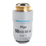
Thermo Fisher Scientific 50X Oil Objective, achromat, coverslip-corrected
1,748,800원
Thermo Fisher Scientific
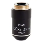
Thermo Fisher Scientific EVOS 100X Oil Objective, achromat, coverslip-corrected
969,700원
배송/결제/교환/반품 안내
배송 정보
| 기본 배송비 |
| 교환/반품 배송비 |
|
|---|---|---|---|
| 착불 배송비 |
| ||
| 교환/반품 배송비 |
| ||
결제 및 환불 안내
| 결제수단 |
|
|---|---|
| 취소 |
|
| 반품 |
|
| 환급 |
|
교환 및 반품 접수
| 교환 및 반품 접수 기한 |
|
|---|---|
| 교환 및 반품 접수가 가능한 경우 |
|
| 교환 및 반품 접수가 불가능한 경우 |
|
교환 및 반품 신청
| 교환 절차 |
|
|---|---|
| 반품 절차 |
|