


Thermo Fisher Scientific MUC-1 Monoclonal Antibody (ZM32)
인간 MUC1 단백질 검출용 마우스 단클론 항체(ZM32). 포르말린 고정 파라핀 절편에서 IHC(P) 적용 가능. Ready-to-use 제형으로 편리한 사용. 단백질 A 정제, 4°C 보관, 인체 MUC1 관련 연구에 적합.
- 판매단위
- pk
카탈로그
1개 옵션 · 카탈로그 번호를 클릭하면 복사됩니다Thermo Fisher Scientific · Thermo Fisher Scientific MUC-1 Monoclonal Antibody (ZM32)
Thermo Fisher Scientific MUC-1 Monoclonal Antibody (ZM32)
Applications
- Immunohistochemistry (Paraffin) (IHC (P))
Tested Dilution: Ready-to-use (150–200 µL)
Product Specifications
| 항목 | 내용 |
|---|---|
| Species Reactivity | Human |
| Host / Isotype | Mouse / IgG1, kappa |
| Class | Monoclonal |
| Type | Antibody |
| Clone | ZM32 |
| Immunogen | Human milk-fat globule membranes (HMFGM) |
| Conjugate | Unconjugated |
| Form | Liquid |
| Purification | Protein A |
| Storage Buffer | Tris with BSA, NP-40 |
| Contains | <0.1% sodium azide |
| Storage Conditions | 4°C |
| Shipping Conditions | Wet ice |
Product Specific Information
This product is diluted and provided in a ready-to-use formulation.
A recommended positive control tissue for this product is Cholangiocarcinoma, although other positive controls may also be used.
The primary antibody is intended for laboratory professional use in detecting the corresponding protein in formalin-fixed, paraffin-embedded tissue stained via manual qualitative immunohistochemistry (IHC). It is used following the primary diagnosis of tumor by conventional histopathology.
This monoclonal antibody reacts with MUC1, a large cell surface mucin glycoprotein expressed by glandular and ductal epithelial cells and some hematopoietic lineages. MUC1 is abundantly expressed in lactating mammary glands and overexpressed in >90% of breast carcinomas and metastases. It associates with all four c-erbB receptors and localizes with EGFR in mammary tissue. The MUC1 gene contains seven exons and produces several alternatively spliced variants. The major expressed form is a type I transmembrane protein with a large extracellular tandem repeat domain that is highly O-glycosylated. Alterations in glycosylation are often seen in epithelial cancer cells.
Antibody to EMA is useful as a pan-epithelial marker for detecting early metastatic loci of carcinoma in bone marrow or liver.
For optimal staining, pretreatment of deparaffinized tissue with heat-induced or enzymatic epitope retrieval is recommended.
IHC staining allows visualization of antigens through sequential application of primary and secondary antibodies, enzyme complex, and chromogenic substrate. The visible reaction product is interpreted under a light microscope for differential diagnosis.
A positive tissue control must be included in every staining procedure. Positive controls should be properly prepared and processed tissues showing weak positive staining to ensure sensitivity and detect reagent degradation.
Negative control tissues may be internal or external based on organizational guidelines. Negative components should demonstrate absence of specific staining to confirm validity of results.
Target Information
MUC1 (Mucin 1, Episialin, MAM-6, CA 15-3, PEM, EMA) is a large transmembrane mucin glycoprotein expressed by most glandular and ductal epithelial cells and some hematopoietic cell lineages. It contains multiple O-linked glycans and shows high polymorphism, with 30–90 tandem repeats depending on the allele.
MUC1 overexpression, aberrant localization, and altered glycosylation are associated with carcinomas. Multiple alternatively spliced transcript variants exist, with some full-length isoforms identified. Transgenic MUC1 associates with all four c-erbB receptors and localizes with EGFR in lactating glands.
For Research Use Only. Not for use in diagnostic procedures. Not for resale without express authorization.
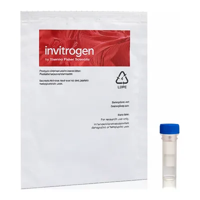
제품 이미지
(이미지 없음 또는 제거됨)
Thermo Fisher Scientific 상품 둘러보기
전체보기
문의
0개 · 배송·재고 문의는 실시간 상담이 빠릅니다아직 등록된 문의가 없어요.