


Thermo Fisher Scientific Cytokeratin, Pan Monoclonal Antibody (ZM374)
Ready-to-use monoclonal antibody (clone ZM374) for IHC detection of pan-cytokeratin in formalin-fixed, paraffin-embedded tissues. Reacts with all 20 cytokeratin subtypes, including CK7, CK8, CK18, and CK20. Suitable for broad epithelial and carcinoma i...
- 판매단위
- pk
카탈로그
1개 옵션 · 카탈로그 번호를 클릭하면 복사됩니다Thermo Fisher Scientific · Thermo Fisher Scientific Cytokeratin, Pan Monoclonal Antibody (ZM374)
Applications
- Immunohistochemistry (Paraffin) (IHC (P))
Tested Dilution
- Ready-to-use (150–200 µL)
Product Specifications
| 항목 | 내용 |
|---|---|
| Host / Isotype | Mouse / IgG |
| Class | Monoclonal |
| Type | Antibody |
| Clone | ZM374 |
| Immunogen | Various human cytokeratin peptide fragments |
| Conjugate | Unconjugated |
| Form | Liquid |
| Storage Conditions | 2–8°C |
| Shipping Conditions | Wet ice |
Product Specific Information
This product is diluted and in a ready-to-use formulation.
A recommended positive control tissue for this product is Pancreas, however positive controls are not limited to this tissue type.
The primary antibody is intended for laboratory professional use in the detection of the corresponding protein in formalin-fixed, paraffin-embedded tissue stained in manual qualitative immunohistochemistry (IHC) testing. It is intended for use after the primary diagnosis of tumor has been made by conventional histopathology using non-immunological histochemical stains.
Anti-cytokeratin clone ZM374 is a mixture of anti-cytokeratin antibodies reacting with all 20 subtypes of cytokeratins. Unlike pan-cytokeratin clone AE1/AE3, ZM374 reacts with CK7, CK8, CK18, and CK20.
In normal tissues, ZM374 is reactive with most epithelial types, including bile ducts, hepatocytes, bladder epithelium, breast ducts, bronchial epithelium, endometrium, intestinal epithelium (stomach, duodenum, ileum, colon, rectum), pancreas, ovarian epithelium, pancreatic acini, pituitary acini, pneumocytes, prostate, thyroid, skin (positive on basal layer, negative on superficial layers of squamous epithelium), and apocrine and sweat glands.
In tumors, ZM374 is reactive with most carcinomas, including breast, transitional cell (TCC), renal cell (RCC), lung adenocarcinoma, lung small cell, lung squamous cell, endometrial, prostate, ovarian, hepatocellular (HCC), colorectal, stomach, and thyroid carcinomas.
It is negative in certain normal tissues, including brain, lymphocytes, hematolymphoid cells, muscle, nerves, endothelium, and in certain tumors such as most melanomas, sarcomas, lymphomas, primitive neuroectodermal tumors (PNET)/Ewing’s, and gastrointestinal stromal tumors (GIST). Positivity may be seen on some dendritic cells in lymph nodes, endothelia, and muscle cells.
Antibody is used with formalin-fixed and paraffin-embedded sections. Pretreatment of deparaffinized tissue with heat-induced or enzymatic epitope retrieval is recommended.
Immunohistochemical staining techniques allow visualization of antigens via sequential application of a primary antibody, a secondary link antibody, enzyme complex, and chromogenic substrate with washing steps. The enzymatic activation of the chromogen results in a visible reaction product at the antigen site, interpreted using a light microscope to aid in differential diagnosis.
Positive and negative control tissues are essential for proper validation.
- External Positive Control: Fresh autopsy/biopsy/surgical specimens fixed and processed similarly to patient samples; ideally with low levels of target antigen for quality control.
- Negative Control: Internal or external tissues showing absence of specific staining to verify background and specificity.
Target Information
Cytokeratin pan belongs to a subfamily of intermediate filament proteins found in human epithelial tissues, represented by at least 20 different polypeptides (molecular weight 40–68 kDa, pI 4.9–7.8).
Cytokeratin subtype expression patterns vary by epithelium type and differentiation stage, and are valuable in distinguishing epithelial malignancies.
Cytokeratin antibodies assist in tumor differential diagnosis using IHC, cytopathology, and flow cytometry. Their expression varies with cell type, differentiation stage, growth conditions, and disease state. Keratins are widely used as markers in cancer research and tumor diagnostics.
For Research Use Only. Not for use in diagnostic procedures. Not for resale without express authorization.
Thermo Fisher Scientific 상품 둘러보기
전체보기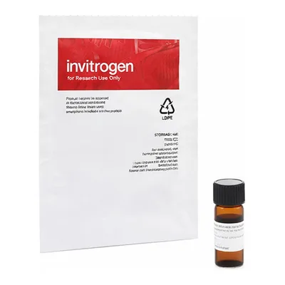

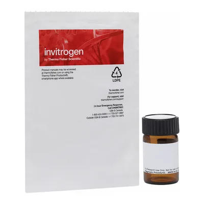
문의
0개 · 배송·재고 문의는 실시간 상담이 빠릅니다아직 등록된 문의가 없어요.