

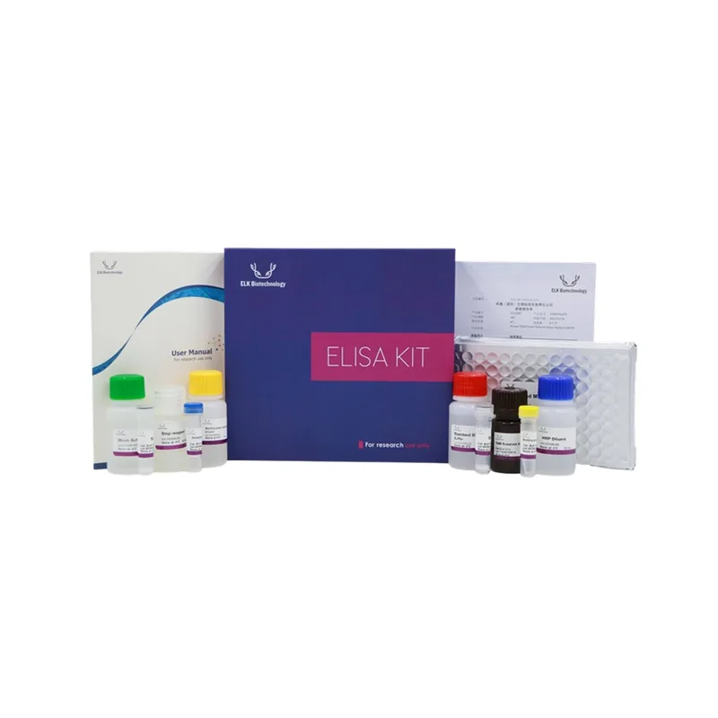
ELK Biotechnology Villin rabbit pAb
Villin 단백질을 인식하는 rabbit polyclonal antibody로, WB 및 ELISA에 적합합니다. 세포골격 및 미세융모 관련 연구에 활용 가능하며, 높은 특이성과 안정성을 제공합니다. -20°C에서 1년간 보관 가능합니다.
- 판매단위
- pk
카탈로그
2개 옵션 · 카탈로그 번호를 클릭하면 복사됩니다ELK Biotechnology · ELK Biotechnology Villin rabbit pAb
제품명
Villin rabbit pAb
기본 정보
| 항목 | 내용 |
|---|---|
| Alternative Names | villin 1 |
| Applications | WB; ELISA |
| Recommended Dilutions | WB 1:500–2000, ELISA 1:10000–20000 |
| Immunogen | Synthesized peptide derived from Villin at AA range: 601–650 |
| Host | Rabbit |
| Storage | -20°C / 1 year |
| Clonality | Polyclonal |
| Isotype | IgG |
| Concentration | 1 mg/ml |
| Observed Band | 90 kD |
| GeneID (Human) | 7429 |
| Human Swiss-Prot No | P09327 |
| Species Reactivity | Human; Mouse; Rat |
세포 내 위치
Cytoplasm, cytoskeleton. Cell projection, lamellipodium. Cell projection, ruffle. Cell projection, microvillus. Cell projection, filopodium tip. Cell projection, filopodium.
Relocalized in the tip of cellular protrusions and filipodial extensions upon infection with S. flexneri in primary intestinal epithelial cells (IEC) and in the tail-like structures forming the actin comets of S. flexneri.
Redistributed to the leading edge of hepatocyte growth factor (HGF)-induced lamellipodia (By similarity). Rapidly redistributed to ruffles and lamellipodia structures in response to autotaxin, lysophosphatidic acid (LPA), and epidermal growth factor (EGF) treatment.
배경 정보
This gene encodes a member of a family of calcium-regulated actin-binding proteins. This protein represents a dominant part of the brush border cytoskeleton which functions in the capping, severing, and bundling of actin filaments. Two mRNAs of 2.7 kb and 3.5 kb have been observed; they result from utilization of alternate poly-adenylation signals present in the terminal exon. [provided by RefSeq, Jul 2008]
제품 이미지
(이미지 없음)
ELK Biotechnology 상품 둘러보기
전체보기
문의
0개 · 배송·재고 문의는 실시간 상담이 빠릅니다아직 등록된 문의가 없어요.